Figure 1.
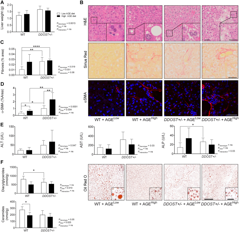
A diet high in AGE content is a promoter of portal vein fibrosis following up-regulation of DDOST+/− in the absence of steatosis. (A) Cull liver weight. (B) Representative H & E stained paraffin-embedded liver tissue sections. (C) Hepatic fibrillar collagen quantification of Sirius Red stained images (left). Representative images of fibrosis stained images of Sirius Red localised around fibrillar fibrosis extending between hepatocytes (right). (D) Hepatic α-SMA positive quantification of immunofluorescent stained images (left). Representative images of α-SMA positive staining extending between hepatocytes (right). (E) Liver function tests for ALT (left), AST (middle) and ALP (right). (F) Hepatic diacylglyceride (top) and ceramide (bottom) accumulation. Representative images of Oil Red O staining for lipid droplets in liver tissue (right). Data represented as means ± SD (n = 4–9/group). *P < 0.05, student’s t-test. Genotype effect P < 0.05, (diet effect) P < 0.05, 2-way ANOVA and multiple comparison of genotype, diet and interaction by Bonferroni’s post hoc test. Representative images scale bar = 50 µm (outside box) and 20µm (inside box).

