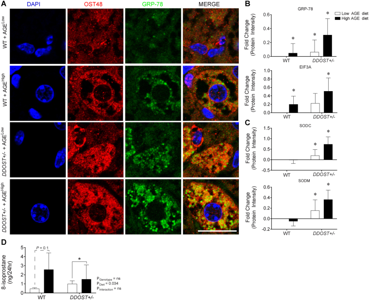
Figure 4.
A high AGE diet promotes liver fibrosis and correlates with increased ER stress markers following increases in DDOST+/− mice. (A) Immunofluorescence for OST48 (red), the ER stress marker GRP-78 (green) on paraffin liver sections (hepatocytes). (B) SWATH-MS protein intensities of ER stress pathway related proteins. GRP78 (top) and EIF3A (bottom). (C) SWATH-MS protein intensities of oxidative stress pathway related proteins. SODC (top) and SODM (bottom). (D) ELISA identifying content of urinary 8-isoprostane. Data represented as means ± SD (n = 4–9/group). *P < 0.05, MSstatsV3.5.1 determined significant log fold changes in the protein intensities between the selected experimental group and the WT low AGE diet group. Representative images scale bar = 20 µm.

