
Fig. 4.
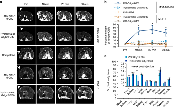
Contrast enhanced MRI with ZD2-Gd3N@C80 of MDA-MB-231 and MCF-7 tumours in mice. a Representative axial T 1-weighted 2D spin-echo MRI images of MDA-MB-231 and MCF-7 tumours in mice. Images were acquired before and at 10, 20 and 30 min after injection of ZD2-Gd3N@C80 and hydroxylated Gd3N@C80 at a dose of 1.67 µmol, or a mixture of 25 µmol kg−1 free ZD2 and 1.67 µmol ZD2-Gd3N@C80 (competitive group). Tumour locations are indicated by white arrow heads. b Analysis of percentage increase of tumour contrast-to-noise ratio (CNR) from images acquired in groups indicated in (a) (data are presented as mean ± s.e.m. n = 4 for MDA-MB-231 tumours and n = 3 for MCF-7 tumours. *P < 0.05 for comparison of the increased CNR ratio of ZD2-Gd3N@C80 in MDA-MB-231 group with that in all the other groups). c Gd biodistribution at 1 week after injection of ZD2-Gd3N@C80 or hydroxylated Gd3N@C80 in MDA-MB-231 tumour models. There was no statistical difference between retention of the contrast agents in all the tested tissues (data are presented as mean ± s.e.m. n = 3)