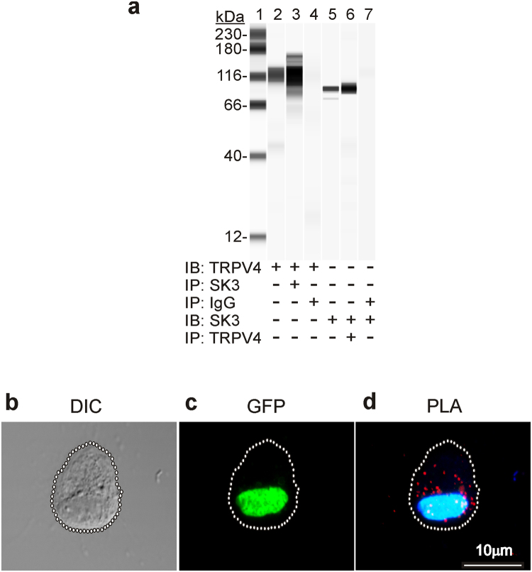
Figure 8.
Co-immunoprecipitation and proximity ligation assay of TRPV4 and SK3 proteins. Immunoblotting (IB) for TRPV4 or SK3 in SK3 or TRPV4 immunoprecipitates (IP) of detrusor membrane fraction (n = 4, a). Lane 1, protein standards; lane 2, IB for TRPV4 in detrusor membrane fraction (1 μg); lane 3 IB for TRPV4 in SK3 IP; lane 4, IB for TRPV4 in non-immune rabbit IgG IP of detrusor membrane fraction; lane 5, IB for SK3 in detrusor membrane fraction (1 μg); lane 6, IB for SK3 in TRPV4 IP; lane 7, IB for SK3 in non-immune rabbit IgG IP of detrusor membrane fraction (n = 4, b). Isolated detrusor PDGFRα+ cell morphology visualized by DIC microscopy (c). The same cell verified as PDGFRα+ by GFP expression. PLA analysis of the same cell using goat anti-TRPV4 and rabbit anti-SK3 antibodies (d).

