. 2017 May 4;8(7):5067–5077. doi: 10.1039/c7sc01182b

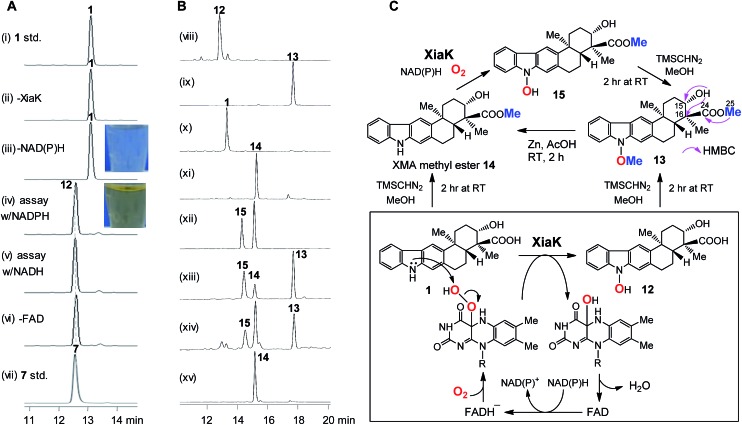
Fig. 3. The in vitro characterization of XiaK. (A) HPLC analysis of the in vitro XiaK enzyme activity assays. A typical XiaK assay was performed in 50 mM Na2HPO4–NaH2PO4 buffer (pH 8.0) for 2 h at 30 °C, comprising 300 μM XMA (1), 1 mM NADPH (or NADH), 1 mM FAD and 5 μM XiaK: (i) XMA (1) standard; (ii) XMA (1) + FAD + NADPH; (iii) XMA (1) + XiaK + FAD; (iv) XMA (1) + XiaK + FAD + NADPH; (v) XMA (1) + XiaK + FAD + NADH; (vi) XMA (1) + XiaK + NADPH; (vii) OXM (7) standard. (B) HPLC analysis of reactions with TMSCHN2. (viii) 12 std; (ix) 12 incubated with TMSCHN2 for 2 h; (x) 1 std; (xi) 1 incubated with TMSCHN2 for 2 h; (xii) an enzyme assay comprising 5 μM XiaK, 0.5 mM 14 and 1 mM NADPH; (xiii) organic extracts from (xii) incubated with TMSCHN2 for 2 h; (xiv) the reaction solution from (xiii) was heated for 2 h, and the conversion of 15 to 14 was observed; (xv) the conversion of 13 to 14 via zinc reduction. (C) Scheme for the chemical transformations of XMA (1) and 12 mediated by XiaK or TMSCHN2.

Fig. 3