Figure 1.

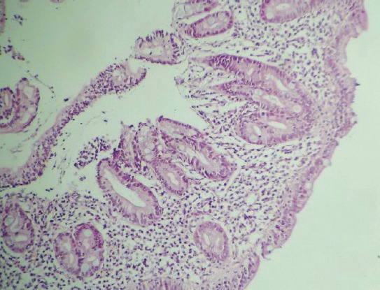
Histopathological examination of the biopsy material of the patients revealed villous atrophy, increase in the intraepithelial number of lymphocytes, and cryptic hyperplasia.

Histopathological examination of the biopsy material of the patients revealed villous atrophy, increase in the intraepithelial number of lymphocytes, and cryptic hyperplasia.