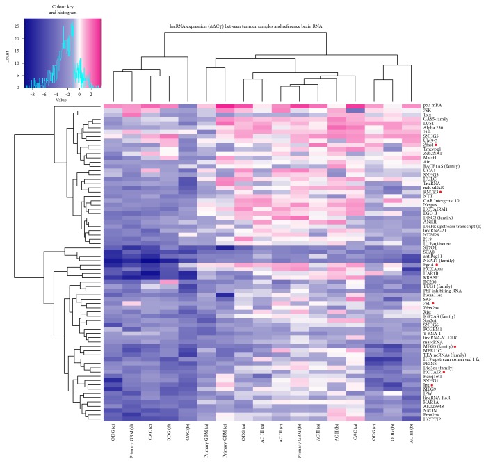
Figure 1.
Heat map of lncRNA profiling analyses of glioma samples. The figure represents ∆∆CT values of differentially expressed lncRNAs in glioma samples compared to human brain reference RNA. Data are presented on a colour scale where shades of blue represent decreased expression and pink as increased expression, with ∆∆CT cut-off values set at −1 and 1. On the top of the figure is presented unsupervised Pearson's hierarchical clustering of samples. AC: astrocytoma of WHO grade II or III; OAC: oligoastrocytoma; ODG: oligodendroglioma. Genes denoted by red dot were selected for further qPCR validation and analysis.

