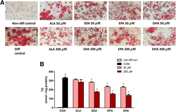
Fig. 3.

Effect of ω-3 fatty acids on TG accumulation in 3T3-L1 adipocytes. Two-day post-confluency preadipocytes were incubated with differentiation medium in the presence of ALA, SDA, EPA, and DHA (0, 50, and 200 μM) for 6 days. a Morphological observation and Oil Red O staining of 3T3-L1 cells photographed using a microscope (X200). Lipid droplets were stained in red. b Quantification of intracellular TG content. Data were obtained from three independent experiments. Absorbance value is given as mean ± SD; different from non-diff control cells: # P < 0.05; different from diff control cells: * P < 0.05
