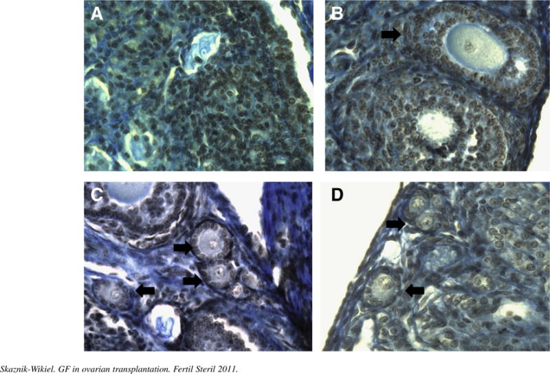
FIGURE 3.

Histologic sections illustrate the appearance of a grafted frozen and thawed ovary in different treatment groups (arrows indicate resting and early growing follicles). (A) Vascular endothelial growth factor (VEGF). (B) Vehicle. (C) VEGF + granulocyte colony-stimulating factor (G-CSF). (D) G-CSF + stem cell factor (SCF). Notice the absence of primordial follicles in VEGF-only and vehicle-treated groups.
