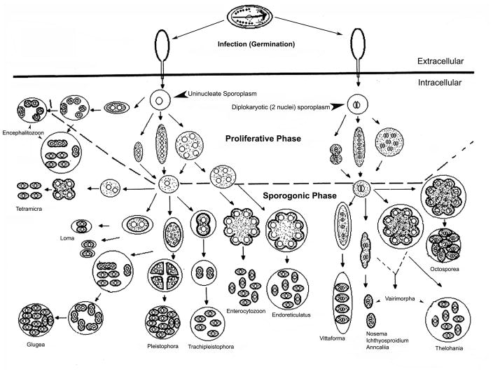
Figure one. Microsporidian Life Cycles.
The initial phase of infection involves spores being exposed to the proper environmental conditions that cause germination of the spores and polar tube extrusion. The polar tube pierces the plasma membrane (solid black line) of the host cell, and the sporoplasm travels through the polar tube into the host cell. The sporoplasm then divides during the proliferative phase and the morphology of this division is used for determination of microsporidian genera. The sporoplasm on the left is uninucleate, and the cells that are produced from it represent the developmental patterns of several microsporidia with isolated nuclei. The sporoplasm on the right is diplokaryotic and it similarly produces the various diplokaryotic developmental patterns. Cells containing either type of nucleation will produce one of three basic developmental forms. Some cycles have cells that divide immediately after karyokinesis by binary fission (e.g. Anncaliia). A second type forms elongated moniliform multinucleate cells that divide by multiple fission (e.g. some Nosema species). The third type forms rounded plasmodial multinucleate cells that divide by plasmotomy (e.g. Endoreticulatus species). Cells may repeat their division cycles one to several times in the proliferative phase. The intracellular stages in this phase are usually in direct contact with the host cell cytoplasm or closely abutted to the host ER; however, the proliferative cells of Encephalitozoon (and probably Tetramicra) are surrounded by a host formed parasitophorous vacuole throughout their development, and the proliferative plasmodium of the genus Pleistophora is surrounded by a thick layer of parasite secretions that becomes the sporophorous vesicle in the sporogonic phase. The sporogonic phase is illustrated below the dashed line. Some of the microsporidian genera maintain direct contact with the host cell cytoplasm during sporogony; i.e. Nosema, Ichthyosporidium, Anncaliia, Enterocyotozoon and probably Tetramicra. The remaining genera form a sporophorous vesicle as illustrated by the circles around developing sporogonial stages. It should be noted that in the Thelohania cycle and the Thelohania-like part of the Vairimorpha cycle, the diplokarya separate and continue their development as cells with isolated nuclei. Adapted with permission from Wittner M and Weiss LM. (Eds.) The Microsporidia and Microsporidiosis. Washington, DC: ASM Press. 1999 (70).

