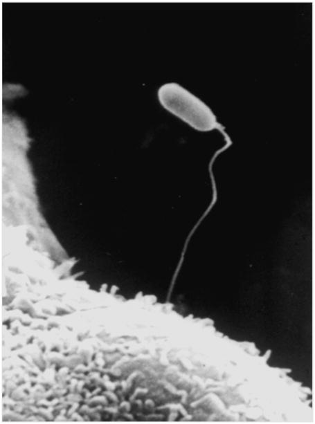
Figure three. Scanning electron micrograph of microsporidia infection of a host cell.
Scanning electron micrograph of extruded polar tube of a spore of Encephalitozoon intestinalis piercing and infecting Vero E6 green monkey kidney cells in tissue culture. Reprinted with permission from Wittner M and Weiss LM. (Eds.) The Microsporidia and Microsporidiosis. Washington, DC: ASM Press. 1999 (70) and with the kind permission of Kock, N.P., C. Schmetz, J. Schottelius, Bernhard Nocht Institute for Tropical Medicine, Hamburg, Germany; published in Kock N.P. 1998. Diagnosis of human pathogen microsporidia (dissertation).

