
Figure 5.
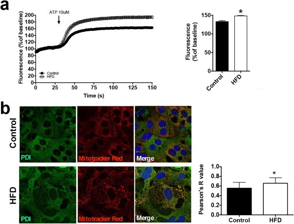
HFD hepatocytes have enhanced mitochondrial Ca2+ signals and increased ER–mitochondrial colocalization. (A) ATP‐induced mitochondrial Ca2+ signals were measured in hepatocytes isolated from mice chronically fed an HFD for 6 months and their chow control littermates (n = 10 cells from three mice). The histogram shows the peak amplitude of mitochondrial Ca2+ signals at 150 seconds. (B) Labeling of ER (PDI, green) and mitochondria (Mitotracker Red, red) indicates an increase in the interaction of both organelles in HFD hepatocytes; colocalization test of immunostaining images (n = 3). Statistical significance was determined using a Student t test. All error bars represent the SEM. *P < 0.05.