
Figure 6.
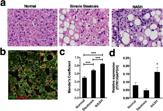
Increased association of ER and mitochondria in human fatty liver disease. (A) Representative images of hematoxylin and eosin staining of a normal, simple steatosis, and NASH human liver biopsy. Regions of lipid accumulation (negative image) are present in both simple steatosis and NASH. In addition to steatosis, NASH specimens also display inflammatory infiltrate and fibrosis. Scale bar = 20 μm. (B) Representative image of a histologically normal human liver biopsy specimen stained with markers for an ER protein (PDI, green) and a mitochondrial protein (Tom‐22, red). Colocalized pixels are highlighted in white. The inset shows a magnified area within the field. Scale bar = 20 μm. (C) Mander's colocalization coefficients of mitochondria to ER shows that the fraction of mitochondria associated with ER is increased in both simple steatosis (n = 5 patients) and NASH (n = 4) in comparison with normal liver (n = 5) and that the coefficient for biopsies from NASH patients is significantly greater than that for patients with simple steatosis. Coefficient values are based on 25 separate determinations in each biopsy specimen. (D) InsP3R1 mRNA expression in human liver specimens is significantly increased in NASH patients compared with control and simple steatosis (n = 6 biopsies per condition). Statistical significance was determined by way of analysis of variance (C) or by using a Student t test (D). All error bars represent the SEM. *P < 0.05. ***P < 0.001.