Figure 1.

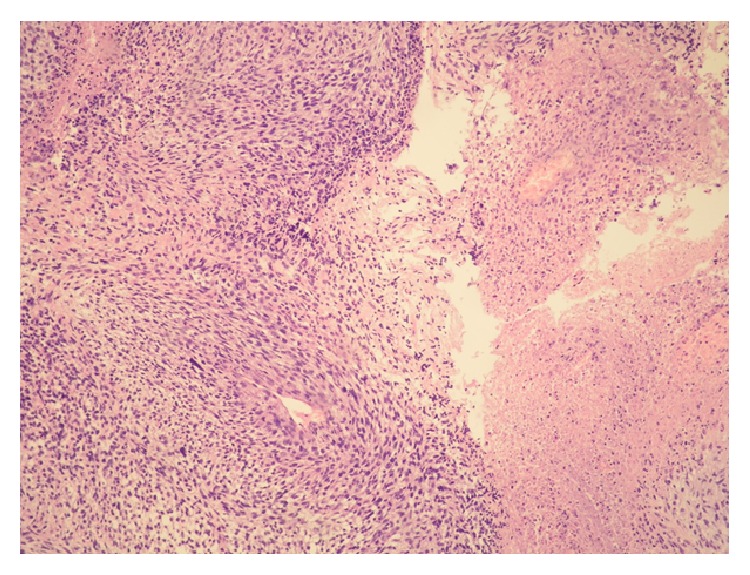
Malignant peripheral nerve sheath tumor histologically showing fascicles of spindle cells (left) and necrosis (right) (HE, ×100).

Malignant peripheral nerve sheath tumor histologically showing fascicles of spindle cells (left) and necrosis (right) (HE, ×100).