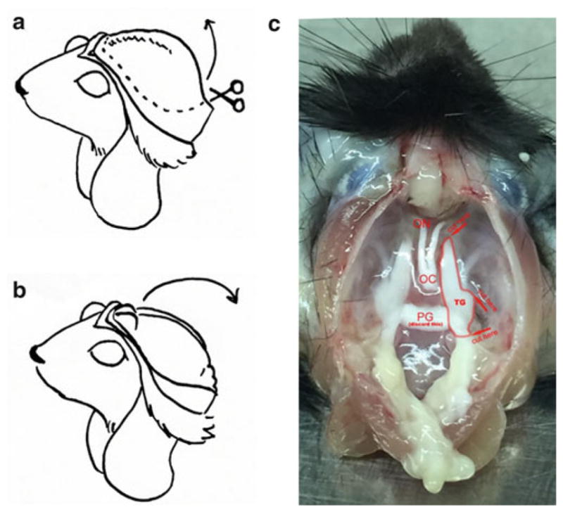
Fig. 3.

TG isolation. After separating the head from the carcass, cut the skin from neck to between the eyes, and fold the skin flaps under the mouse’s chin (a). Cut the exposed skull along left and right, to between the eyes, then lift the bone up and away (a). Pull the brain out toward you (b), exposing the base of the skull (c). TG dissection: Picture shows a mouse head after brain removal. Trigeminal ganglion is double lined. Arrows indicate cutting sites and the orientation of the cutting. Abbreviations: TG trigeminal ganglion, ON optic nerve, OC optic chiasm, PG pituitary gland. The optic nerves and chiasm should be visible at the top, the TGs along the base to either side, and the pituitary gland along the base between the TGs. Cut the connective tissue anchoring the TGs to the skull, and sever the nerve where it enters the skull—you should now be able to lift out the TGs (C)
