
Figure 6. Blood breakdown products induce regulated necrosis in cortical neurons by recruiting both ferroptotic and necroptotic cell death mechanisms.
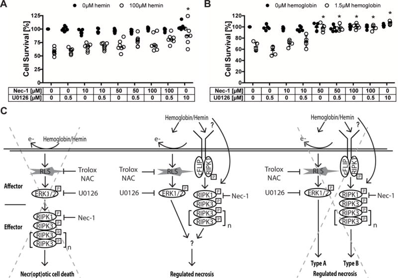
(A–B) Concentration-responses of Necrostatin-1 cotreated with U0126 (sub-threshold dose) in hemin (A) and hemoglobin (B) toxicity. Values represent medians for hemin and mean ± SD for hemoglobin. * p<0.05 versus hemin or hemoglobin alone. (C) Three alternative hypotheses suggesting how hemoglobin or its breakdown product hemin induces cell death with ultrastructural features of necrosis: (1) Hemoglobin or hemin generate reactive lipid species (RLS), which increase phospho-ERK1/2. Phospho-ERK1/2 induces RIP1 that executes necroptotic cell death via RIP3. In this case, hemorrhagic stroke would induce ferroptosis as the affector phase of death leading to a necroptotic effector phase. (2) Hemoglobin or hemin induces necroptotic (RIP1/3) and ferroptotic (phospho-ERK) cell death mechanisms independently and these then converge at a yet to be identified common denominator into a necrotic morphology. (3) Hemoglobin or hemin induces necroptotic (RIP1/3) and ferroptotic (phospho-ERK) cell death mechanisms that independently lead to different types of regulated necrosis. Our data supports the second hypothesis. NAC – N-acetylcysteine, Nec-1 – Necrostatin-1.