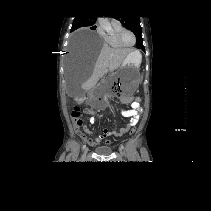
Figure 3.

Coronal view of contrast-enhanced CT demonstrating fluid and air collection in the pancreatic region consistent with infected pseudocyst (down arrow) and low-density subcapsular hepatic fluid collection consistent with hepatic abscess (right arrow).
