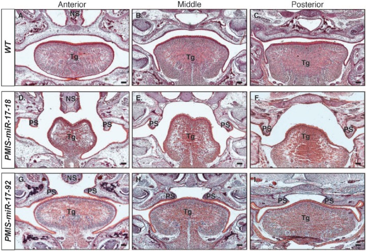
Figure 2.
Histologic analyses show distinct patterns of clefting at embryonic day 18.5 in PMIS-miR-17-18 and PMIS-miR-17-92 embryos. Hematoxylin and eosin staining of embryonic day 18.5 embryos shows differences in cleft palate defects observed in PMIS-miR-17-18 and PMIS-miR-17-92 embryos. (A–C) Anterior, middle, and posterior coronal sections of wild-type (WT) embryos show complete fusion of the palate. (D–F) Coronal sections of the PMIS-miR-17-18 embryos shows that arrest in palatogenesis occurs before elevation of the palatal shelves. (G–I) Coronal sections of PMIS-miR-17-92 embryos shows that arrest in palatogenesis occurs after elevation of the palate shelves but before extension and fusion at the midline. Scale bar = 100 μm. NS, nasal septum; PS, palatal shelves; Tg, tongue.

