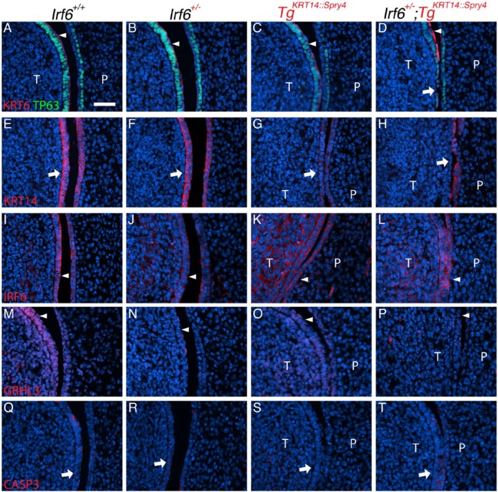
Figure 3.
Irf6 and Spry4 interaction leads to loss of periderm and oral adhesions between the tongue and palate at E13.5. (A–T) Images and analysis of oral adhesions and immunostaining to examine expression of KRT6/TP63 (A–D), KRT14 (E–H), IRF6 (I–L), GRHL3 (M–P), and activated caspase 3 (CASP3, Q–T) between the tongue and palate. Genotypes separated by columns, including wild-type (A, E, I, M, Q), Irf6 heterozygous (Irf6+/–) (B, F, J, N, R), TgKRT14::Spry4 (C, G, K, O, S), and Irf6+/–;TgKRT14::Spry4 embryos (D, H, L, P, T). (A–D) Immunostaining showed an abnormal expression pattern of both KRT6 and TP63 (compare white arrowheads in A–D), with loss of periderm and basal cells (white arrow) in Irf6+/–;TgKRT14::Spry4 embryos (D). (E–H) KRT14 immunostaining highlighted adhesions in TgKRT14::Spry4 embryos (G) and loss of lingual oral epithelium in Irf6+/–;TgKRT14::Spry4 embryos (H). (I–L) IRF6 stained both the periderm and basal epithelial cells in wild-type embryos (I) and Irf6 heterozygous (J). There appeared to be ectopic IRF6 expression in the lingual mesenchyme of both TgKRT14::Spry4 (K) and Irf6+/–;TgKRT14::Spry4 (L) embryos. Epithelial IRF6 expression did not appear different (I–L, white arrowheads). (M–P) GRHL3 expression was found in both the periderm and basal epithelial cells of tongue and, to a lesser extent, the palatal shelves (M). There was reduced GRHL3 expression in the periderm of Irf6 heterozygous (N) and TgKRT14::Spry4 embryos (O). Loss of periderm contributed to the apparent reduction GRHL3 expression in Irf6+/–;TgKRT14::Spry4 (P) embryos (white arrowheads). (Q–T) Activated caspase 3 expression appeared more prominent in sites of oral adhesion in Irf6+/–;TgKRT14::Spry4 (T) embryos (white arrows). Scale bar: (A–T) 40 µm. P, palate; T, tongue.

