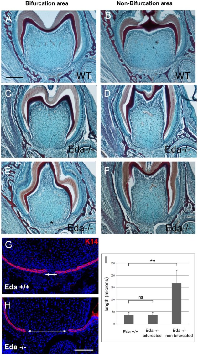
Figure 3.
Hertwig’s epithelial root sheath (HERS) extension defects in Eda mutant mice at postnatal day 7. (A–F) Trichrome-stained frontal sections in the center of the tooth (bifurcation region; A, C, E) and outside the normal furcation region (B, D, F) in the lower first molar. (A, B) Wild type (WT). (C–F) Eda mutants. *Point where the 2 sides of the HERS have almost met in the mutant. (G, H) Keratin 14 expression in the bifurcation region in WT (G) and mutant (H) HERSs. Arrows indicate gap between the lingual and buccal sides of the HERS. (I) Graph showing Eda mutants can be divided into 2 groups dependent on the distance between the 2 sides of the HERS at the center of the tooth. Scale in A–F: 200 μm. G, H: 100 μm. *P < 0.05. **P < 0.01. Values are presented as mean ± SD. ns, not significant.

