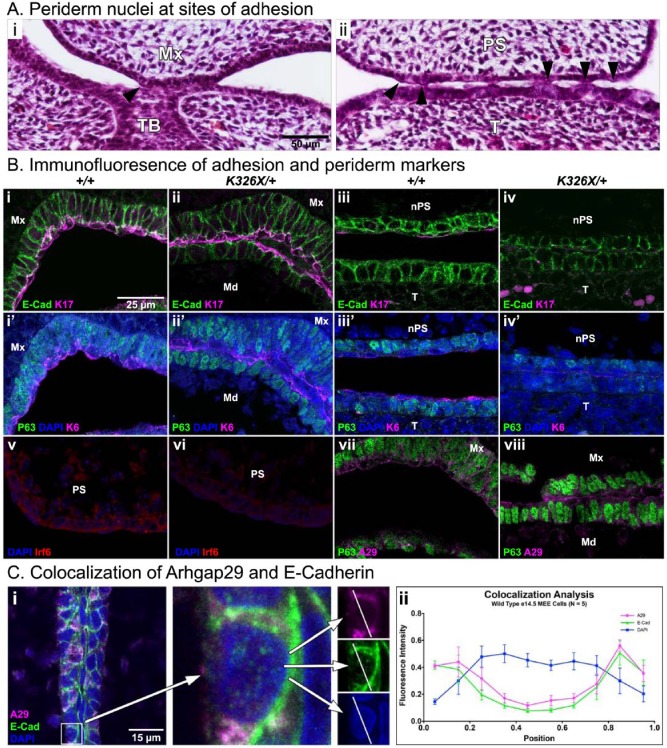
Figure 4.
Characterization of the oral periderm at sites of adhesion. (A, B) Histological view of e14.5 Arhgap29K326X/+ oral adhesions. Arrowheads indicate periderm nuclei present at points of contact between the maxilla and mandibular tooth bud (A, i) and the palatal shelf and tongue (A, ii). (B) Immunofluorescent staining for adhesion and periderm markers in paired littermates (genotypes designated in columns). Primary antibodies are E-cadherin (i-iv, green), keratin 17 (i-iv, magenta), p63 (i′-iv′, v-viii, green), keratin 6 (i′-ii′, magenta), Irf6 (v-vi, red), and Arhgap29 (vii-viii, magenta). i′-iv′ are sections adjacent to i-iv. Note the presence of keratin 17–positive, keratin 6–positive, and p63-negative cells (periderm) at the site of mandibular (Md) and maxillary (Mx) epithelial contact in i, i′, ii, and ii′. At an adhesion involving the nasal side of the palatal shelf (nPS) and the tongue (T; iii-iv, iii′-iv′, location the same as Fig. 2B, arrowheads), E-cadherin is present, while keratin 17 and keratin 6 expression is barely detectable. (v-vi) Irf6 is present in oral epithelium and periderm with bright speckles in nuclei, as previously described (Richardson et al. 2009). (vii-viii) Arhgap29 is expressed in the oral epithelium and the periderm, with strongest signal occurring in periderm cells at the ends of adherent spans (viii). (C) Colocalization analysis for E-cadherin and Arhgap29 at the medial edge epithelium (MEE). On a single z-plane, lines were drawn across the center of an epithelial cell (i). The fluorescence intensity along these lines was compared for each channel. Resulting values for Arhgap29 and E-cadherin were significantly correlated (P < 0.0001, Pearson correlation). Raw values were normalized and binned into discrete lengths for graphical representation (ii). Mx, maxilla; Md, mandible; TB, tooth bud; PS, palatal shelf; nPS, nasal side of palatal shelf; T, tongue.

