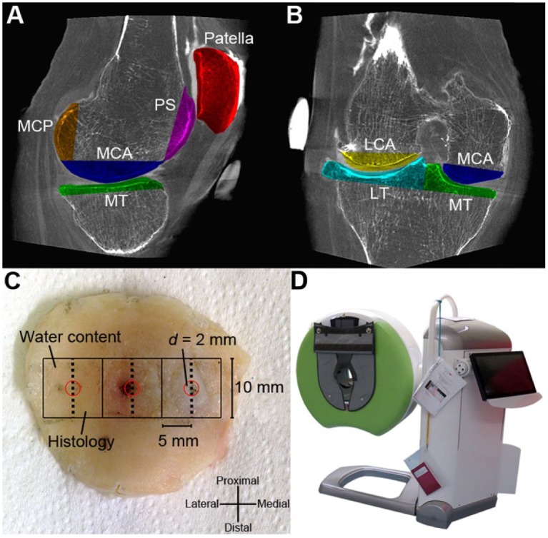
Figure 1.

Excised joint surface pieces (n = 7) in sagittal (A) and coronal (B) planes (PS = patellar surface, LCA = lateral condyle anterior part, MCA = medial condyle anterior part, LT = lateral tibia, MT = medial tibia, MCP = medial condyle posterior part). (C) Illustration of the patella with indentation sites marked (d = 2 mm, red circles). A 1 × 1 cm2, full-thickness piece was harvested symmetrically around each indentation site, and was subsequently halved through the indentation site for water content determination and histology (0.5 × 1 cm2 each). (D) A clinical peripheral cone beam computed tomography (CBCT) scanner (Verity, Planmed Oy, Helsinki, Finland) used in this study.
