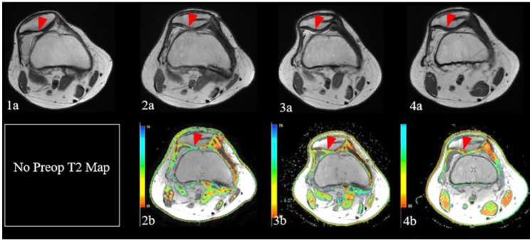
Figure 3.
Sequential magnetic resonance images demonstrate chondral defect over the lateral patellar facet on initial fast spin echo proton density (FSE PD) weighted images (1a, red arrowhead), in the setting of lateral patellar subluxation. Subsequent FSE PD image (2a) following patellar particulated juvenile allograft cartilage (PJAC) transplantation, medial patellofemoral ligament (MPFL) reconstruction, and tibial tubercle osteotomy demonstrates restoration of normal patellofemoral alignment, as well as good fill of the chondral defect by hyperintense repair tissue (red arrowhead), with corresponding prolongation of relaxation times and lack of normal chondral stratification on corresponding T2 map (2b, red arrowhead). FSE PD image (3a) obtained approximately 1 year following repair demonstrates decreased relative hyperintensity of the graft, with corresponding decrease in T2 relaxation values and early chondral stratification on T2 mapping (3b, red arrowhead), indicative of ongoing graft maturation. Follow up images obtained approximately 2.5 years following the initial surgery demonstrate further normalization of graft signal on FSE PD weighted images (4a, red arrowhead), with further decrease in relaxation times and progressive stratification of repair tissue on T2 maps (4b, red arrowhead), reflecting further graft maturation.

