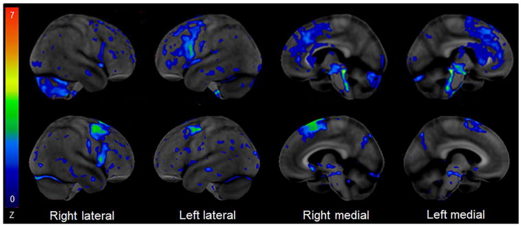
Figure 2.
[18F] Fluorodeoxyglucose PET scan using the Cortex Suite software reveals mild hypometabolism of the left posterior frontal cortex, bilateral supplemental motor cortices, midbrain, superior cerebellar peduncle and right cerebellum in a case of Richardson’s syndrome (top row) and mild hypometabolism in bilateral posterior frontal cortices, and right supplemental motor cortex in a patient with primary progressive apraxia of speech (bottom row) suggestive of an underlying primary tauopathy.

