
Figure 6.
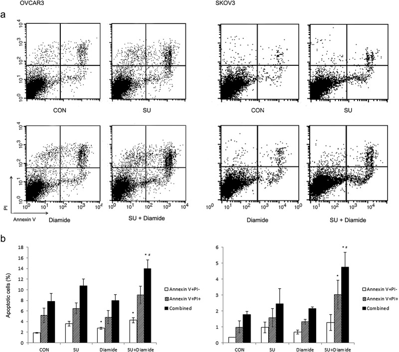
Enhancement of sulforaphane-induced cell death by diamide. OVCAR3 and SKOV3 cells were pretreated with diamide (50 or 100 μM) for 1 h before treatment with 6.25 μM sulforaphane (SU). Cells were stained with Annexin V and propidium iodide (PI). Apoptotic cells were detected using flow cytometry. (A) Representative density plots of dual annexin/PI staining are shown. (B) The percentage of apoptotic cells is shown. Lower right quadrant (annexin V-positive and PI-negative) indicates early apoptotic cells. Upper right quadrant (annexin V and PI-positive) represents necrotic or late apoptotic cells. Both early and late apoptotic cells (Combined) were calculated as the incidence of apoptotic cell death. Values are mean ± SE, n = 3. An asterisk (*) indicates a significant difference (p < 0.05) in the cell death rate compared to vehicle control and a hash sign (#) indicates a significant difference (p < 0.05) compared to treatment with diamide.