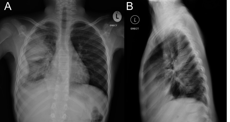
Figure 1.
Chest X-ray in a 9-year-old girl with fever not responding to antibiotics. (A) Anteroposterior radiograph demonstrates lobulated density with a convex intrathoracic margin along the lateral aspect of the chest with a broad base in keeping with pleural pathology. This is continuous with the lamellar effusion extending to the apex. In addition, there is a well-defined elliptical density in the mid portion of the right chest and no air-bronchogram within it consistent with a ‘pseudotumour’. (B) Lateral radiograph serves to confirm that the elliptical density lies within the major fissure and that it is also pleural based.

