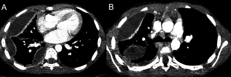
Figure 2.
Non-sequential CT scan slices in the same patient as (figure 1A). (A) A more cranial slice at the level of the carina confirms that there is a right-sided effusion with a loculated component and that in addition the pseudotumour on chest X-ray is indeed a loculated collection in the major fissure. The lateral loculation demonstrates a thick enhancing medial edge consistent with an empyema. (B) A more caudal slice at the level of the cardiac ventricular chambers demonstrates that the pleural collection is multiloculated with lateral and medial components adjacent to the spine and heart, all demonstrating enhancing inner margins. This was important for making management decisions. No air-space consolidation was demonstrated to suggest an underlying pneumonia on lung windows (not shown here).

