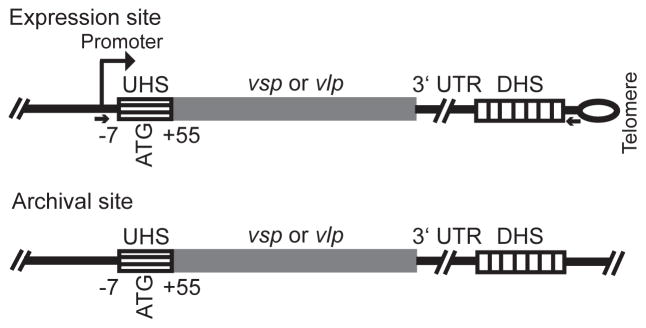
Figure 1.
Schematic representation of expression and archival sites for vsp and vlp genes of Borrelia hermsii. The drawing is not to scale. The locations of the UHS regions that surround the start codon (ATG), the 3′ untranslated region (UTR), and DHS elements are given. By the numbering system, +1 is the transcriptional start position at the expression site. The UHS regions at the archival sites varied in length to the extent that they were >90% identical to the UHS at the expression site. The expression site is adjacent to a hair-pin telomere, indicated by the loop. The small arrows give the location of PCR primers (see text) for amplification of the expression site but not the silent site vsp or vlp gene.

