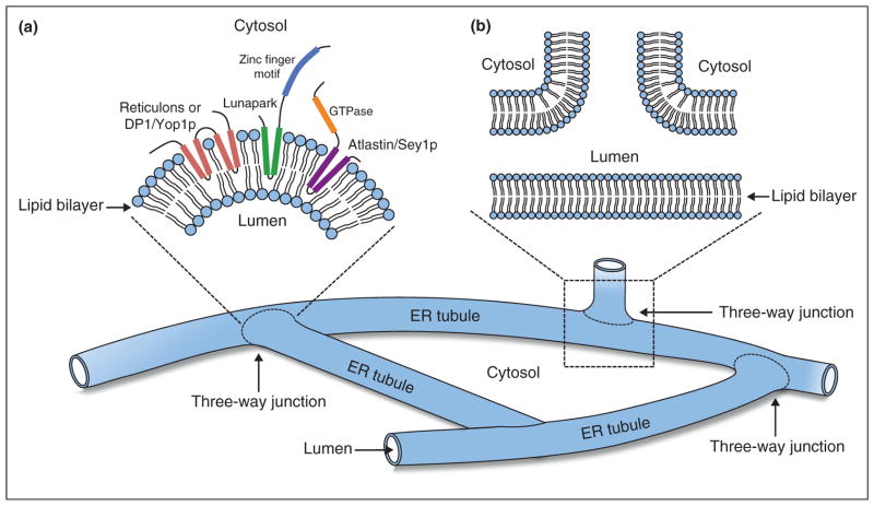
Figure 2.
Schematic diagram of the ER shaping proteins at three-way junctions. (a) Membrane topology around a three-way junction where reticulons (coral), DP1/Yop1p (coral), Lunapark (green and blue) and atlastin (purple and orange) insert into the outer leaflet of the phospholipid bilayer from the cytosolic side of the membrane. (b) A cross-section view of a three-way junction.

