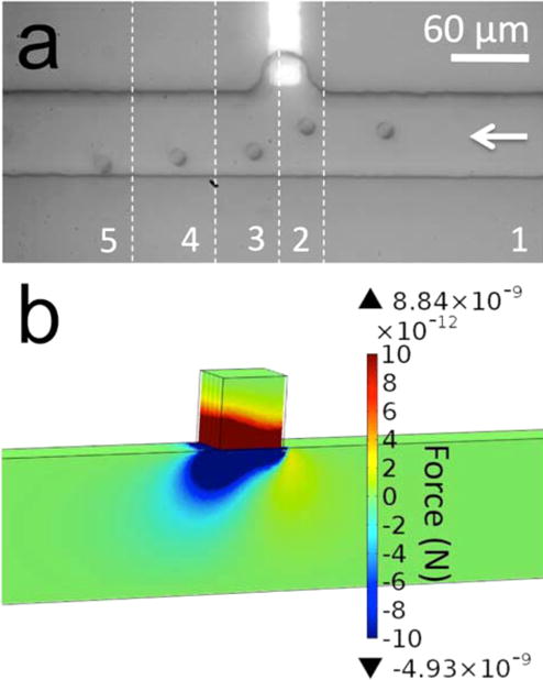
Figure 1.

(a) A series of optical micrographs, which show nDEP repulsion of a B-cell from the BPE tip under AC-only electric field in Tris DEP buffer. Each image slice (numbered sequentially 1–5) is separated by 2.5 s. ERMS,avg = 5.7 kV/m (t = 0 s) to 17.7 kV/m (t = 5s). (b) Simulated magnitude of the y-component of FDEP in the xy-plane at z = 5 μm.
