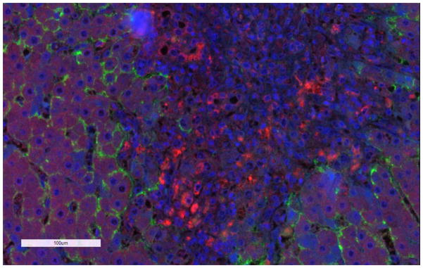
Figure 1. Immunofluorescence staining of MRP1 in a liver tissue section from a patient with primary biliary cirrhosis.
Dual staining of MRP1 (red) and Na+/K+ ATPase (green), which was used as a basolateral membrane marker. The nuclei are stained in blue. The general methods are described in Pomozi et al.201 MRP1 and Na+/K+ ATPase antibodies were purchased from Abcam and SantaCruz Biotechnology, respectively.

