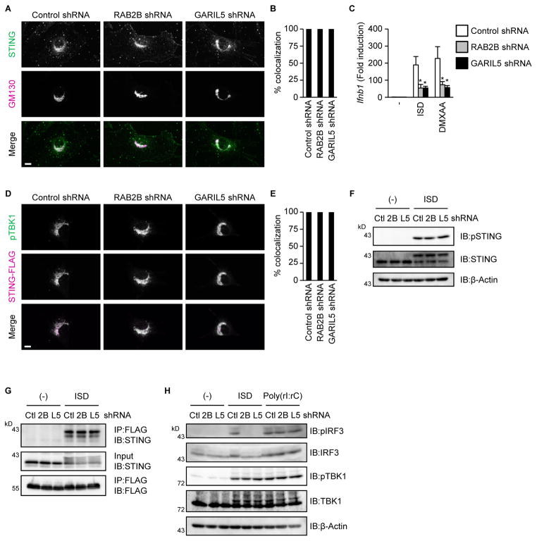
Figure 4. RAB2B-GARIL5 complex regulates the phosphorylation of IRF3.
(A and B) Primary MEFs stably expressing the indicated shRNAs were established by retroviral transduction and stimulated with DMXAA (100 μg/ml) for 1 h. Colocalization of endogenous STING and GM130 was observed under a fluorescence microscope (A) (scale bars, 10 μm). The frequency of colocalization of endogenous STING with GM130 in DMXAA-stimulated MEFs was determined (B). The graph shows means ± SD (n = 3); *P < 0.01.
(C) Primary MEFs stably expressing the indicated shRNAs were stimulated with ISD (1000 ng/ml) or DMXAA (100 μg/ml) for 1 h. The levels of Ifnb1 mRNA were measured by quantitative RT-PCR. The results shown are means ± SD (n = 3); *P < 0.01.
(D and E) Primary MEFs stably expressing the indicated shRNAs, together with STING-FLAG, were stimulated with DMXAA (100 μg/ml) for 1 h. The localization of phosphorylated TBK1 and STING-FLAG was observed by a fluorescence microscope (D) (scale bars, 10 μm). The frequency of colocalization of phosphorylated TBK1 with STING-FLAG in DMXAA-stimulated MEFs was determined (E). The graph represents means ± SD (n = 3); *P < 0.01.
(F) Primary MEFs stably expressing the indicated shRNAs were stimulated with ISD (1000 ng/ml) for 1 h. Whole cell lysates were subjected to immunoblot analysis using anti-phospho STING, anti-STING, and anti-β-actin.
(G) Primary MEFs stably expressing the indicated shRNAs, together with STING-MYC and FLAG-IRF3-S385A/S386A, were stimulated with ISD (1000 ng/ml) for 1 h. Whole cell lysates were immunoprecipitated and immunoblotted with the indicated antibodies.
(H) Primary MEFs stably expressing the indicated shRNAs were stimulated with ISD (1000 ng/ml) or poly (rI:rC) (1000 ng/ml) for 1 h. Whole cell lysates were subjected to immunoblot analysis using anti-phospho IRF3, anti-IRF3, anti-phospho TBK1, anti-TBK1, and anti-β-actin.

