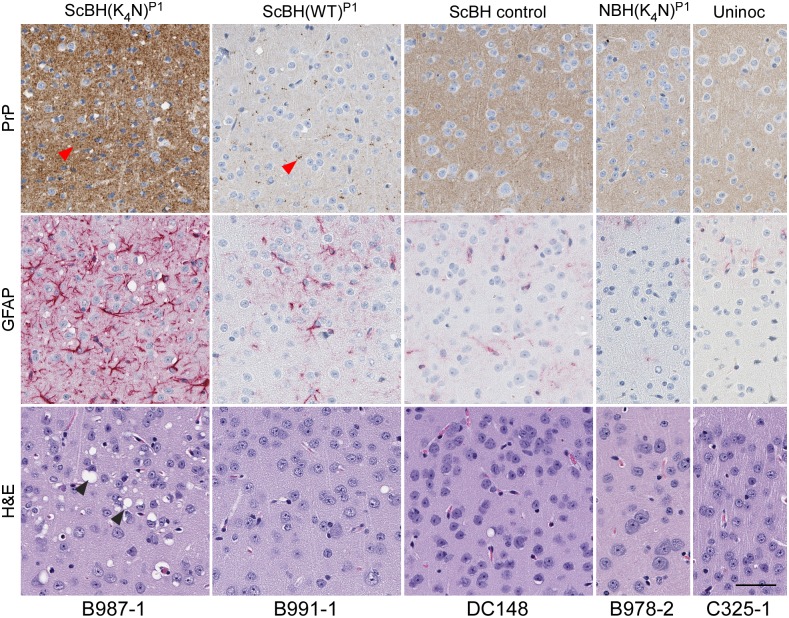
Fig 7. Histopathology of cerebral cortex of Tg7 ScBH(WT or K4N)P1 mice and controls.
Slides were stained using a PrP antibody (EP1802Y), GFAP antibody (for astrocytic activation), or hematoxylin and eosin. Animal numbers are displayed below images. Red arrow heads denote PrP aggregates and black arrows denote vacuolization (spongiosis). Scale bar indicates 50 microns.

