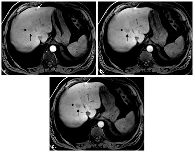
Figure 3.
A 49-year-old man with hepatic adenomas. Three postcontrast images obtained at 3:58 (a), 13:49 (b), and 19:58 (c) after gadoxetate injection show progressive hepatic parenchymal enhancement and vessel clearance. The first image set (a) was rated an inadequate hepatocyte phase (average reader grade = 1.0, SIRLV = 1.18), whereas the second (b) and third (c) image sets were rated adequate (average reader grade = 2.0 and 2.0; SIRLV = 1.47 and 1.73, respectively). Note minimal improvement in lesion conspicuity between image set (b), obtained at 13:49, and set (c), obtained at 19:58 postcontrast injection. SIR, signal intensity ratio; LV liver/vein.

