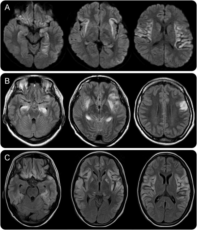
Figure 1. MRI lesions in the acute stage of cryptogenic new-onset refractory status epilepticus.
Initial brain MRI at the onset of status epilepticus is unremarkable, but a few days later, MRI shows symmetric increased diffusion-weighted images (DWIs) or T2/fluid-attenuated inversion recovery (FLAIR) signals in the hippocampus, amygdala, insula, claustrum, thalamus, perisylvian operculum, and basal ganglia (A–C). These newly appearing lesions are likely associated with persistent seizure activity that was highly refractory to conventional antiepileptic treatments. Brain MRIs were obtained on day 20 of the onset of status epilepticus (A, patient 3), day 3 (B, patient 6), and day 74 (C, patient 9). (A) DWI and (B and C) FLAIR images.

