
Figure 1. Hierarchical assembly of the embryonic nerve ring.
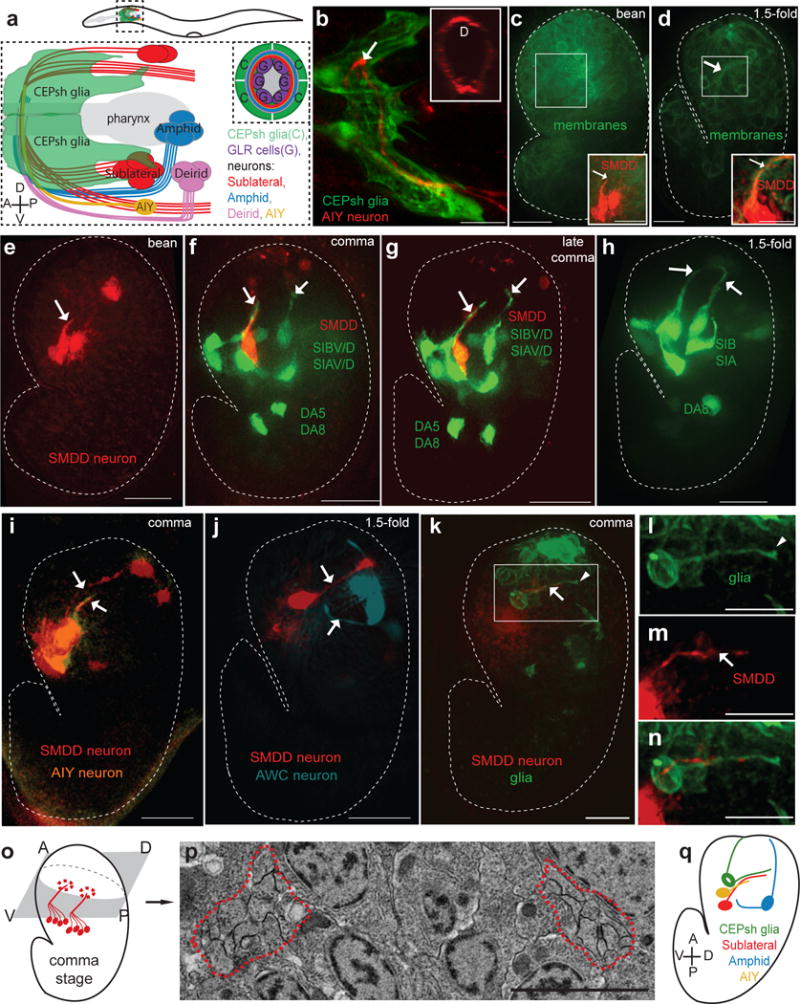
(a–b) The post-embryonic nerve ring is populated by neuronal commissures and enveloped by CEPsh glia. (a) Schematics of lateral view of post-embryonic nerve ring. (b) Imaging of post-embryonic nerve ring in L4 animals. Inset, cross-sectional view. Pttx-3::mCherry (AIY, red), Phlh-17::myristoylated-GFP (CEPsh glia). (c–n) Formation of the embryonic nerve ring starts at late bean stage, with early entry of sublateral commissure axons and CEPsh glia and later entry of other components. (c,d) Phis-72:: myristoylated-GFP (cell membranes). (c–k) Dotted line: embryo outline. D: dorsal, V: ventral, A: anterior, P: posterior. Arrow: axon, arrowhead: CEPsh glia. Scale bars: 10μm. (c-g,i-k,m,n) Pttx-3::mCherry (SMDD, red; AIY, pseudocolored orange in (I). (e–h) Pceh-17::GFP, sublateral neurons SIAV/D, SIBV/D. (j) Phlh-16::GFP (SIA/SIB, red; AWC, pseudocolored blue). (k–n) Pmir-228:: myristoylated-GFP (glia). (b–n) Expression patterns are described in Supplementary Methods and Supplementary Tables S8, S10. l–n: magnified view of boxed region in k. (o,p) A single pair of bilateral bundles are observed in electron micrographs of early comma stage embryos. Schematic (o) and magnification (p) of electron micrograph section. The magnified region corresponds to the region outlined in the blue box of Supplementary Fig. S1g. Red outline: axon bundle. Scale bar: 2μm. (q) Summary of imaging results. CEPsh glia and pioneer neurons enter NR path first, followed by amphid and AIY neurons.