
Figure 2. CEPsh glia and SubL axons functionally pioneer the NR.
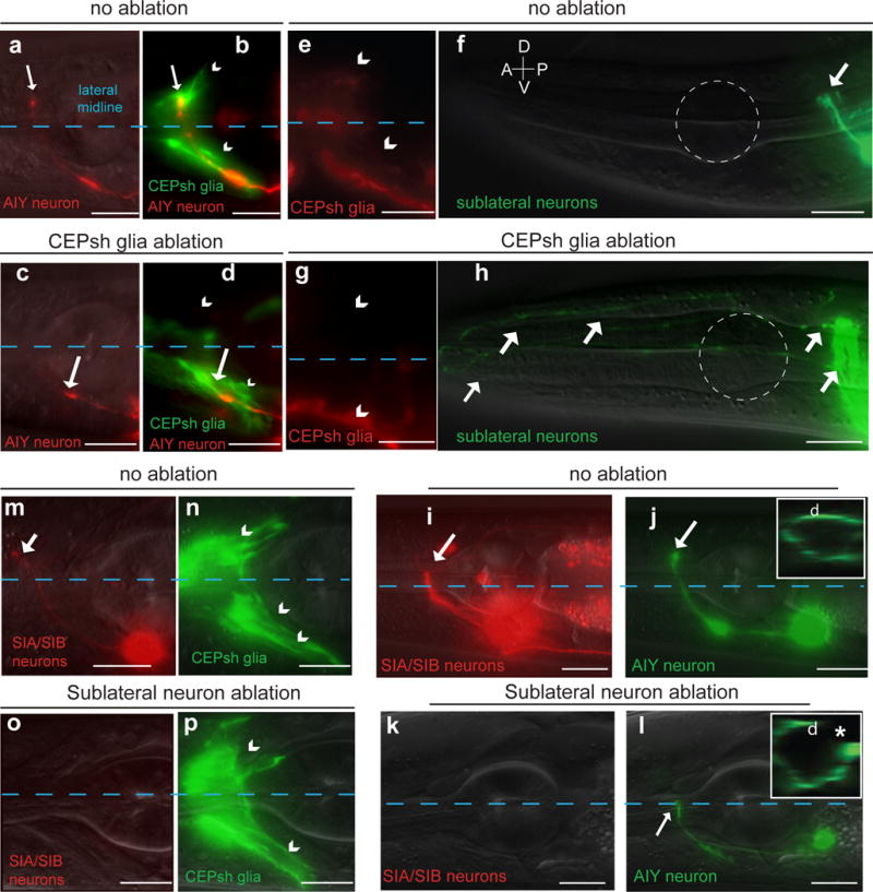
Nerve-ring entry of AIY follower axons and fasciculation of sublateral commissure axons is abnormal in CEPsh-ablated animals. Ablation of neurons of the sublateral commissure results in abnormal AIY nerve-ring entry but spares CEPsh membrane growth. Scale bars: 10 μm. (a–d) Pttx-3::mCherry (AIY), Pmir-228::GFP (glia). (e–h) Pceh-24::GFP (SIA/SIB/SMD), Pptr-10::RFP (glia). Dotted white outline: 1st pharyngeal bulb. (i–p) Pceh-17::RFP (SIA/SIB), Pttx-3::GFP (AIY, green) or Phlh-17:: myristoylated-GFP (CEPsh glia, green). Reporter expression patterns are described in Supplementary Methods and Supplementary Tables S8, S10. Asterisk: NR axon gap.