
Figure 3. NR axon entry is disrupted in kpc-1; chin-1 mutants.
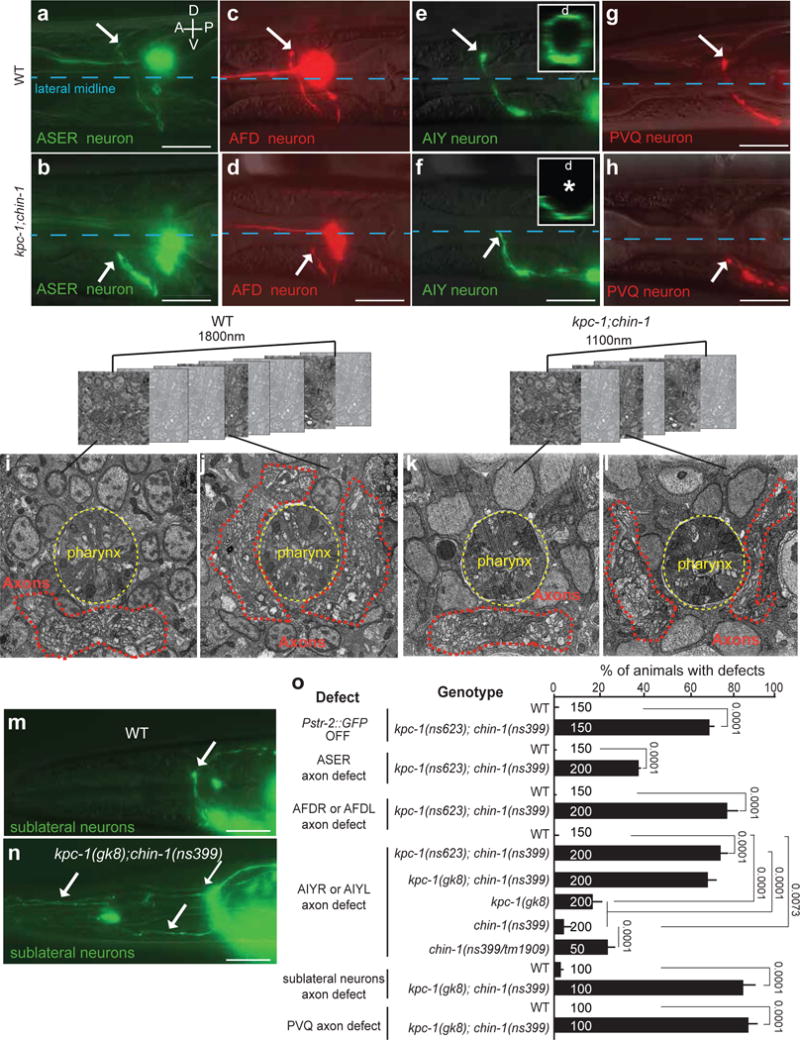
(a–h, m–o) Nerve-ring guidance of axons of different neuron subtypes, and in different commissures, is abnormal in kpc-1; chin-1 mutants. Asterisk, arrow, scale bars: 10μm. D: dorsal, V: ventral, A: anterior, P: posterior. (a,b) Pgcy-5::GFP. (c,d) Pttx-1::RFP. (e,f) Pttx-3::GFP. (b,d,f) kpc-1(ns623); chin-1(ns399). (g,h) Pnpr-11::RFP. (i–l) Nerve-ring structure of L1 animals is abnormal in kpc-1; chin-1 mutants compared to wild-type animals. FIBSEM images of WT (i,j) and mutant (k,l) NR region of age-matched L1 animals. Dotted red line: axons. (h,k,l,n) kpc-1(gk8); chin-1(ns399). (m,n,o) Fasciculation of sublateral commissure neurons is abnormal kpc-1; chin-1 L1 mutant animals compared to wild-type animals. Pceh-24::GFP. (a–h, m–n) Reporter expression patterns are described in Supplementary Methods and Supplementary Tables S8, S10. (o) Numbers inside bars: total animals scored per genotype, n=4 independent scoring experiments. Mean +/− Error bars: SEM. Numbers above bars of significance, p values from Fisher’s exact test. ns: non significant.