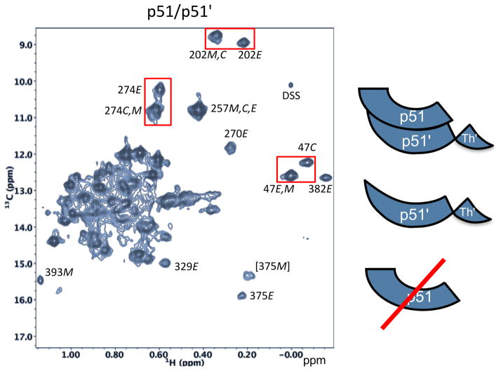
Figure 9. 1H-13C HMQC spectrum of [U-2H,13CH3-Ile]p51.
Species contributing to the spectrum, illustrated in the schematic on the right, include the p51/p51′ homodimer and the p51 monomer. Resonance intensities suggest that the sample is ~ 80% dimeric. Resonances are attributed to the extended conformation (labeled E), the compact (monomer) structure (labeled C), and to the monomer (labeled M) as indicated. Resolved resonances for Ile47 (fingers domain), Ile202 (palm domain), and Ile274 (thumb domain). The spectrum was obtained under conditions that favor dimerization: 800 mM KCl, 20 mM MgCl2. Despite using these conditions, a significant Ile393 resonance attributed to the monomer species is also present.

