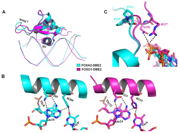
Figure 6.
Comparison with the previous FOXO1-DBD/DBE2 structure (PDB ID: 3CO7). (A) Overall structure comparison between FOXA2-DBD/DBE2 (cyan) and FOXO1-DBD/DBE2 (magenta). (B) Most hydrogen bonding interactions were conserved between the structures. (C) Structure comparison shows that the N-terminal tail of FOXO1-DBD is located closer to the minor groove although the proteins is bound to the same DNA. Arg157 and Asn158 of FOXO1 (magenta) interact with the DNA backbone. The presence of Pro160 and Pro161 of FOXA2 (cyan) orients the N-terminal tail of FOXA2-DBD away from the minor groove.

