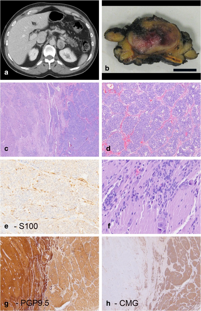
Fig. 1.
Patient 1 was a 57-year-old male who was incidentally discovered to have a left adrenal mass lesion on a CT scan performed for hematuria (a). On cross section, the tumor consisted of a demarcated lesion arising within the adrenal gland (b) (size bar 1 cm). H&E images show a biphasic lesion (c) with nested areas of pheochromocytoma (d) that includes S100 positive sustentacular cells (e). Other areas show neuropil-type stroma with immature small neuroblastic cells and rare ganglion cells (f). PGP9.5 and chromogranin corresponding to the same area seen in panel C show distinct labeling in the two components. The neuroblastoma component is more strongly positive for PGP9.5 (g) while chromogranin (CMG) expression is largely restricted to the pheochromocytoma component of the tumor (h)

