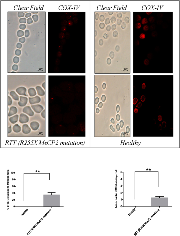
Figure 4.
Identification of mitochondria in mature RBCs of RTT patients by IF. Upper panel: Immunofluorescence microscopy analysis of the RBCs isolated from the RTT patients carrying the R255X MeCP2 mutation (n = 3) (left panel) and from healthy individuals (n = 3) (right panel). RBCs were allowed to adhere to glasses by cytospinning and were probed with an anti COX-IV antibody. RTT RBCs displayed a COX IV+ dotted pattern that was undetectable in healthy RBCs. Clear field acquisition highlighted a normal bi-concave shape of the RBCs. The experiment was carried out in triplicate by using the same blood specimen. Lower panel: the percentage of RBCs displaying at least 1 mitochondria was calculated in 10 different fields (left panel); the average number of mitochondria per RBC was calculated by counting the number of dots for each RBC in the different fields (right panel). The results are the means ± S.E.; **significantly different from control (**p < 0.005, unpaired τ Student’s test).

