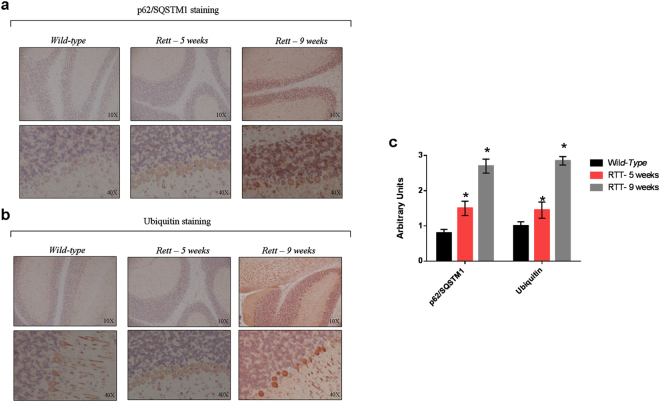
Figure 5.
Accumulation of autophagy reporter substrates in the cerebellum of murine models of RTT. Immunohistochemistry analysis of the cerebellum from 9-weeks-old wild-type mice and 5- (asymptomatic) and 9-weeks (symptomatic) old RTT mice knock-out for MeCP2 (n = 3 for each experimental group). For the three experimental conditions, the organs isolated from the different animals were studied. Slices (n = 4) from each organ were probed with anti-p62/SQSTM1 (a) and an anti-Ub antibody (b). An age-linear increase in the staining for both p62/SQSTM1 and Ub was observed in all the cerebellum layers of RTT mice compared to the cerebellum of wild-type animals. (c) Bar graph showing the semiquantitative evaluation of p62/SQSTM1 and Ub immunoreactivity, expressed as arbitrary unit. The results were expressed as mean number ± S.E., with an interobserver reproducibility of ±95%. The staining of the cerebellum of 5-weeks mice was compared to that of wild-type mice, and the staining of the 9-weeks mice to that of 5-weeks mice. Differences were evaluated by a Student’s τ test and were considered significant at p value ≤ 0.05.

