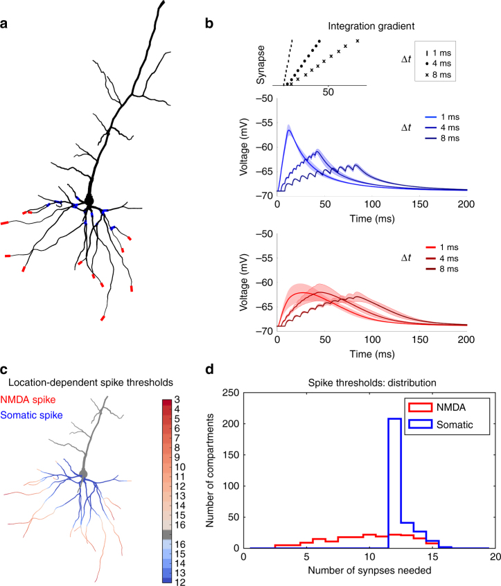
Fig. 1.
Location dependence of local and global regenerative events. a Proximal (blue) and distal (red) locations on a thin basal branch of the detailed neuron model. b Temporal integration gradient along a basal dendrite (reproducing experimental results by Branco and Häusser47). Top panel: activation sequences with different interspike intervals. Middle panel: proximal stimulation requires high temporal coincidence, as the somatic depolarisation drops substantially when inputs are not active synchronously. The solid line is the mean over all proximal compartments shown in panel a, the shaded region is one s.d. Bottom panel: distal stimulation results in similar somatic depolarisation over a bigger range of interspike intervals. The solid line is the mean over all distal compartments shown in panel a, the shaded region is one s.d. c For each dendritic compartment in the neuron model, synapses are activated until either an NMDA spike or an action potential is generated. Stimulating proximal compartments results in an action potential before reaching the threshold for NMDA spikes (blue). Stimulating distal regions results in NMDA spike generation while staying sub-threshold for somatic spikes (red). Intenser colours denote that less synapses are needed to evoke a spike. The numbers next to the colour bar represent the number of synapses needed for a spike. d For each compartment in c, the number of synapses needed to evoke the spikes was stored and represented as a histogram. NMDA spikes (red) generally require less synapses in our model than somatic spikes (blue)

