Figure 5.
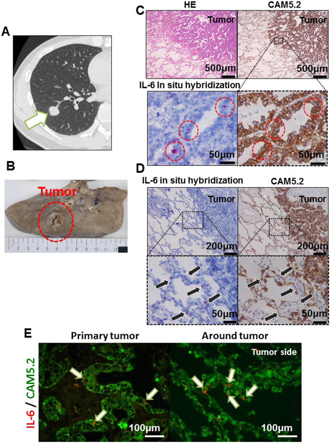
The IL-6 in situ hybridization of the clinical samples. A computed tomography (CT) image (A) and a picture of the surgical specimen (B) of a 52-year-old male patient with EGFR mutation-positive (Exon 19 deletion) lung adenocarcinoma (Case 4, lesion 5 in Table 1). (C,D) The in situ hybridization of IL-6 mRNA and HE and CAM5.2 staining of the cells in the tumor (C) and just around the tumor (D) in lung adenocarcinoma. IL-6 mRNA-positive lung cancer cells were detected in the clinical samples (red circle in C), and some normal lung epithelial cells located immediately around the tumor were also IL-6 mRNA-positive (arrow); however, the normal lung epithelial cells were mostly negative. (E) Double staining of IL-6 mRNA and CAM5.2 in the tumor cells and just around the tumor.

