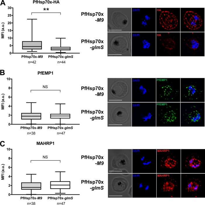
FIG 7 .
Knockdown of PfHsp70x does not inhibit export of PfEMP1 to the host cell. (A to C) PfHsp70x-M9 and PfHsp70x-glmS parasites were fixed with acetone and stained with antibodies against HA, PfEMP1, or MAHRP1. DAPI was used to mark parasite cell nucleus. (Right) From left to right, the images are phase-contrast micrographs of parasites, parasites stained with DAPI, parasites stained with anti-HA antibody or antibody against exported protein, and fluorescence merge image. Representative images are shown. Bars, 5 µm. (Left) The mean fluorescence intensity (MFI) for each protein was calculated for individual cells and shown as box-and-whisker plots, with whiskers representing the maximum and minimum MFI. For HA, the MFI was calculated for the entire infected RBC. For PfEMP1 and MAHRP1, MFI was calculated for the exported fraction only. Significance was determined using an unpaired t test (**, P ≤ 0.01; NS, not significant).

