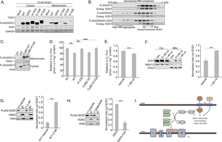
FIG 6.
SOD1-mediated suppression of respiration is upstream of its mitochondrial localization. (A) HEK 293 cells were transfected with Flag-tagged WT, G93A, K122Q, K122R, or K122E SOD1. The cells were lysed, and mitochondria were fractionated from the cytosol. The two fractions were subjected to SDS-PAGE and immunoblotted for Flag-SOD1 present in the mitochondria versus the cytosol. (B) Flag-tagged WT, G93A, or G93A K122E SOD1 was induced with tetracycline for 48 h in T-REx 293 cells. The cells were lysed, and the lysates were then loaded onto a Superdex 200 10/300 GL size exclusion column and were eluted in fractions. The fractions were subjected to SDS-PAGE and immunoblotted for Flag-SOD1 and endogenous SOD1. MM, molecular mass. (C) Flag-tagged WT, CytB2-WT, or CytB2-K122E SOD1 was overexpressed in HEK 293 cells. The cells were lysed, and mitochondria were fractionated from the cytosol. The two fractions were subjected to SDS-PAGE and immunoblotted for Flag-SOD1 present in the mitochondria versus the cytosol. (D) Flag-tagged WT, K122E, CytB2-WT, or CytB2-K122E SOD1 was overexpressed in HEK 293 cells. Oxygen rates were measured with an Oroboros O2K respirometer. The experiment was carried out in triplicate. Asterisks indicate significant differences (**, P < 0.01; ***, P < 0.001); error bars represent SEM. (E) Rotenone (1 nM) or a vehicle was added to HEK 293 cells 30 min prior to measurement of oxygen consumption rates with an Oroboros O2K respirometer. The experiment was carried out in quadruplicate. (F) Rotenone at 1 or 10 nM or a vehicle was added to HEK 293 cells 30 min prior to lysis of the cells and fractionation of the mitochondria from the cytosol as in the experiment for which results are shown in panel A. The fractions were immunoblotted for the presence of endogenous SOD1 in the mitochondria and cytosol. In each of the six replicates, SOD1 levels were first normalized to mitochondrial and cytosolic loading controls and then normalized to the ratio of mitochondrial SOD1 in the 1 nM rotenone-treated sample. (G) Flag-tagged WT-SOD1 was overexpressed in HEK 293 cells. A 1 mM concentration of 3-NP was added for 16 to 18 h before the cells were lysed and mitochondria (M) were fractionated from the cytosol (C). The two fractions were subjected to SDS-PAGE and immunoblotted for Flag-SOD1 present in the mitochondria versus the cytosol. In each of the three replicates, Flag-SOD1 levels were normalized first to mitochondrial and cytosolic loading controls and then to the ratio of mitochondrial Flag-SOD1 in the 3-NP-treated sample. (H) Flag-tagged G93A SOD1 was overexpressed in HEK 293 cells. A 25 mM concentration of galactose in low-glucose (2 mM) medium was added 24 h before the cells were lysed and assayed as in the experiment for which results are shown in panel G. In each of the three replicates, Flag-SOD1 levels were normalized first to mitochondrial and cytosolic loading controls and then to the ratio of mitochondrial Flag-SOD1 in the untreated sample. (I) An Erv1/MIA40 disulfide relay promotes the IMS import of CCS, which in turn promotes the IMS retention of SOD1. The disulfide relay is reset when oxidized cytochrome c accepts an electron from Erv1. OMM, outer mitochondrial membrane; IMM, inner mitochondrial membrane. (Model based on that of Kawamata and Manfredi [41].)

