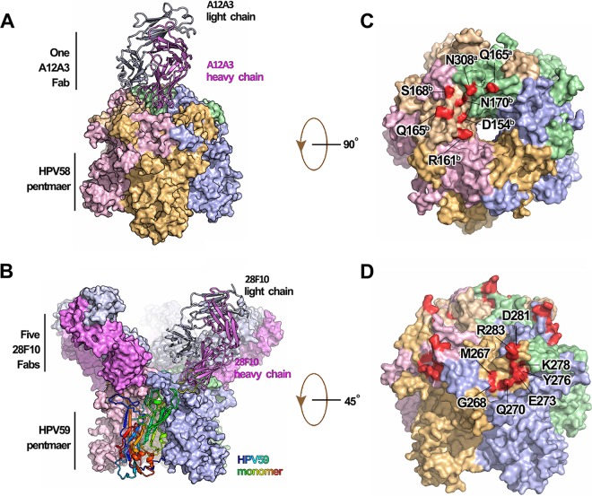
FIG 2 .
Crystal structures of the immune complexes of HPV58p:A12A3 and HPV59p:28F10. (A) HPV58p:A12A3 structure with the Fab shown as a ribbon and the antigen in surface representations. (B) HPV59p:28F10 structure with a monomer and its bound Fab shown as a ribbon and the monomer colored from blue to red (N terminus to C terminus). (C and D) The footprints of MAbs A12A3 (C) and 28F10 (D). Both immune complexes are represented in the same color scheme with different chains: chain a in pale green, chain b in wheat, chain c in light pink, chain d in light orange, and chain e in light blue for the HPV pentamer and the heavy chain in violet and the light chain in white-blue for antibody.

