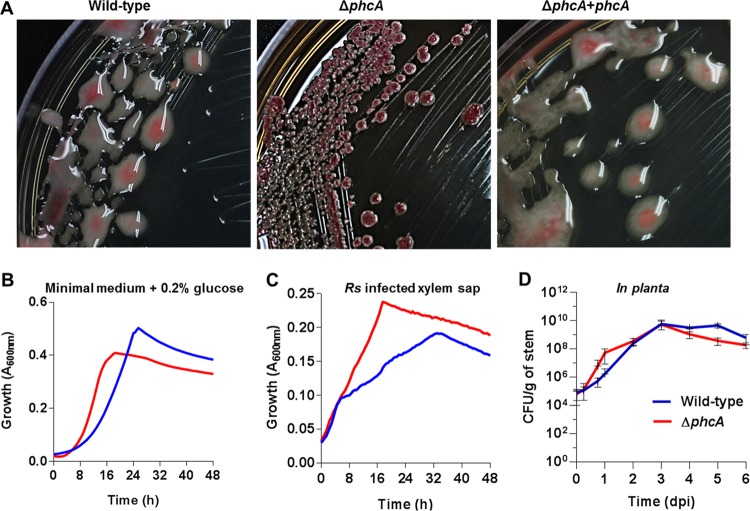
FIG 1 .
Phenotypes of the low-cell-density-mode-mimicking ΔphcA mutant. (A) Colony morphology of R. solanacearum strains on tetrazolium chloride agar plates. Wild-type, phylotype I strain GMI1000; ΔphcA, deletion mutant of GMI1000; ΔphcA+phcA, phcA deletion mutant complemented by inserting one copy of phcA into the chromosome. (B and C) Growth of wild-type R. solanacearum (blue) and of the ΔphcA mutant (red) was monitored over 48 h under aerobic conditions at 28°C in minimal broth supplemented with 0.2% glucose (three biological replicates and four technical replicates, n = 12) (B) or in ex vivo xylem sap harvested from GMI1000-inoculated tomato plants showing early wilt symptoms (four biological replicates, each performed with five technical replicates, n = 20) (C). Each replicate was grown in xylem sap pooled from eight plants. Growth was measured as A600 levels by the use of a BioTek microplate reader. The ΔphcA mutant grew faster than the wild type both in minimal medium (Mann-Whitney test, P = 0.0081) and in xylem sap (analysis of variance [ANOVA], P < 0.0001). (D) Population sizes of the wild-type strain and the ΔphcA mutant in tomato stems following inoculation with 2,000 cells through a cut leaf petiole, as determined by serial dilution plating of ground stem slices. Data represent means of results from nine plants per treatment per time point; bars represent standard deviations.

