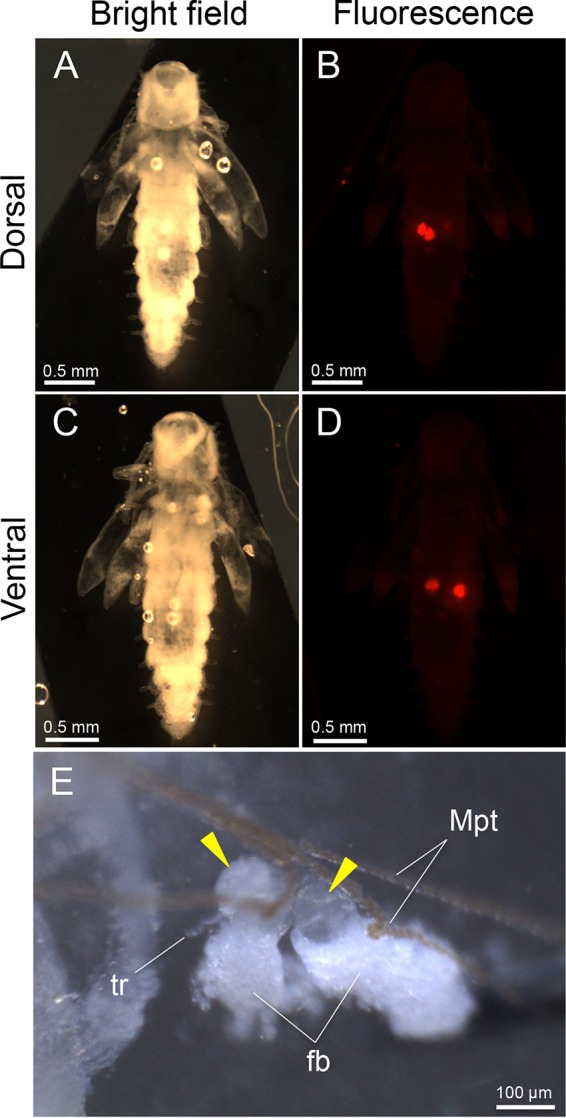
FIG 4 .

Localization and tissue construction of bacteriomes in pupae of O. surinamensis. (A) Bright-field image from the dorsal side. (B) Epifluorescence dissection microscopic image from the dorsal side, in which two dorsal bacteriomes are visualized in red by whole-mount in situ hybridization targeting 16S rRNA of the symbiont. (C) Bright-field image from the ventral side. (D) Epifluorescence dissection microscopic image from the ventral side, in which two ventral bacteriomes are seen in red. In panels A to D, different from Fig. 3F to H, the insect body was not pressed, thereby enabling the observation of the dorsal and ventral bacteriomes separately. (E) Magnified image of two dorsal bacteriomes (arrowheads) dissected from a pupa. Abbreviations: fb, fat body; Mpt, Malpighian tubule; tr, trachea.
